Simon Bertram
EBVS® European Specialist in Veterinary Neurology
DVM MVetMed DipECVN MRCVS
What are congenital vertebral malformations?
Congenital vertebral malformation (CVM) is an umbrella term describing a variety of different birth defects of the vertebral column.
Failure of formation:
-
Butterfly vertebrae - partial or complete failure of formation of the ventral and central portions of the vertebral body
-
Hemivertebrae - failure to form one sagittal half of the vertebrae, including the centrum and neural arch
-
Wedge-shaped vertebrae - incomplete failure to form one sagittal half of the vertebrae with a variable degree of unilateral hypoplasia
-
Transitional vertebrae - vertebra showing features of two adjacent segments of the vertebral column e.g. thoracic and lumbar
-
Articular process dysplasia - aplasia or hypoplasia of the cranial or caudal articular process. See Figure 1
Failure of segmentation:
- Block vertebrae - complete or partial fusion of two adjacent vertebral bodies without an intervertebral disc space between them
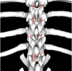 Figure 1: Bilateral/Unilateral articular process aplasia (arrows)
How common are these findings in dogs?
The overall prevalence of CVMs in dogs is not well known but it has been well established to be a very common incidental finding (80-97%) in brachycephalic dogs like English Bulldogs, French Bulldogs and Pugs. These changes can range from just one transitional vertebra to severe complex combinations of multiple CVMs.
The tale of the tail!
Overall, there seems to be a difference between “screw-tailed” brachycephalic breeds like English and French Bulldogs and “non-screw-tailed” brachycephalic breeds like the Pug. Pugs are very predisposed to have transitional vertebra and caudal articular process dysplasia whereas Bulldogs most commonly show butterfly vertebrae, hemivertebrae or wedge-shaped vertebrae with the degree of tail malformations being correlated to these changes. More recently a mutation in the gene DVL2 was linked to thoracic and coccygeal CVMs in Bulldogs but not in Pugs.
When are they relevant?
As stated above these findings are a very common incidental finding and are rarely directly causing any clinical signs (e.g. neck or back pain or varying degrees of myelopathy). The most important tool for deciding if an imaging finding is relevant is to see if it matches the dog’s exact problem. The following gives a rough guideline to help decide if a finding is relevant or not:
Transitional vertebra
- Should be considered incidental.
Caudal articular process dysplasia
-
More likely in Pugs
-
More likely to be clinically relevant if:
-
Bilateral aplasia
-
Between T11 and L1
-
Multiple vertebrae in a row
-
Butterfly vertebrae, hemivertebrae or wedge-shaped vertebrae
- Less common in Pugs but more likely to be clinically relevant if present
- Kyphosis
- A marked degree of Kyphosis secondary to the CVMs is the most reliable marker of relevance.
- A cobb angle over 35º seems to be have the highest sensitivity and specificity.
The clinical picture, treatment and prognosis!
Caudal articular process dysplasia
Clinical signs: Presenting dogs (mainly Pugs) tend to be older (> 7 years) and show a very slowly progressive, non-painful T3-L3 myelopathy. Urinary and faecal incontinence are a common finding.
Pathogenesis: The most common reason for clinical signs are secondary changes compressing the spinal cord. The lack of normal facet joints will cause instability of the spinal cord and can lead to soft tissue bands compressing the spinal cord (sometimes causing CSF accumulation similar to a subarachnoid diverticulum), repetitive micro trauma to the spinal cord and intervertebral disc disease due to increased biomechanical stress.
Diagnosis: Normally a combination of CT and MRI will yield the most information. The CT is superior in assessing the exact underlying CVMs whereas the MRI is superior at assessing the degree of spinal cord compression and ruling out other underlying causes for the clinical signs.
Treatment: Conservative treatment (rest, physiotherapy and low-dose steroids) can be trialled but seems to have a poor long-term outcome. The treatment of choice seems to be surgical stabilisation, with or without spinal cord decompression. Using 3D-printed patient-specific drill guides makes this very safe for the patient. Early research shows that this will be successful in about 75% of dogs.
 Figure 2: Bilateral vertebral stabilisation with bicortical screws and PMMA
Butterfly vertebrae, hemivertebrae or wedge-shaped vertebrae
Clinical signs: Presenting dogs tend to be young (< 12months) and show a slowly progressive, normally non-painful T3-L3 myelopathy. Urinary and faecal incontinence are a common finding.
Pathogenesis: The clinical signs are most commonly caused by a compression of the spinal cord directly by the kyphosis secondary to the CVMs or secondary by intervertebral disc compression. There is a large debate about this process being rather dynamic than static and instability of the vertebral column might also play an important factor.
Diagnosis: Normally a combination of CT and MRI will yield the most information. The CT is superior in assessing the exact underlying CVMs whereas the MRI is superior at assessing the degree of spinal cord compression and ruling out other underlying causes for the clinical signs.
Treatment: Conservative treatment (rest, physiotherapy and low-dose steroids) seems not to be a promising approach. In a study of 13 cases which were managed conservatively 30% where euthanized and 70% slowly progressed. The treatment of choice seems to be an early stabilization of the affected vertebral column with the help of screws and bone cement. Using 3D-printed patient-specific drill guides makes this process very safe for the patients. Preliminary results seem very promising and there is ongoing investigation into the long-term success.
Top Tips
- Congenital vertebral malformations are very common in brachycephalic breeds
- They should be considered incidental if they are detected by chance and cannot be associated with the described clinical signs
- Clinical signs associated with CVMs can be caused by instability or compression
- Treatment of choice is case-dependent vertebral column stabilization with or without spinal cord de-compression