A rescue dog suffering from a severe limb deformity is now back on all four paws and living life to the full after complex surgery.
Yana, a 19-month-old crossbreed who came to the UK from Romania, was walking uncomfortably and was struggling to stand properly when she was referred us.
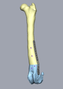
CT scans revealed she had a complex deformity affecting both her tibia and femur of the left pelvic limb, plus a lesser deformity affecting the right pelvic limb.
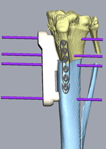
European Veterinary Specialist in Small Animal Surgery Anna Nutt took over Yana’s care, teaming up with Bill Oxley at Vet3D, who produced customised bone guides to enable Anna to carry out corrective surgery.
These patient-specific surgical guide systems are designed using 3D data from CT scans, and enable surgeons to create 3D bone models to plan surgical interventions. In this case, 3D printed bone models, bone cutting guides and reduction guides were then created to enable precise correction of the deformity in the operating theatre.
Yana underwent left femoral and tibial limb straightening and is now enjoying life back with owners Danielle and David Cook in Hemyock, Devon.
Anna said: “Yana’s owners reported that she always seemed stiff when getting up, and that her left hind limb looked bowed. They also noticed that she walked unusually and tended to ‘bunny hop’ on her hind limbs.
“A CT scan of the hind limbs revealed a significant valgus deformity of the left femur and left tibia. In addition, there was mild torsional and also sagittal plane deformity causing a very high tibial plateau angle, the latter of which can predispose to cruciate ligament rupture. Yana also had subluxation of the left hip, likely as a result of the abnormal limb loading.
“We worked with Bill Oxley at Vet3D, and determined that Yana would require an ostectomy of both the distal femur and proximal tibia to correct the deformities and give her the best chance of normal limb function.
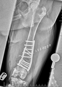
“We operated on Yana and performed a distal femoral ostectomy to correct the valgus, which was reduced and stabilised with a lateral 3.5mm DFO plate and a craniomedial 2.7mm LCP plate.
“We then performed a proximal tibial ostectomy, which was reduced and stabilised with a 3.5mm broad TPLO plate and a 2.7mm cranial LCP plate.
“The tibial ostectomy corrected both the tibial valgus and reduced the tibial plateau angle to six degrees. Post-operative radiographs showed successful bone straightening and good implant positioning.”
Following the operation, Yana was sent home with strict instructions for cage rest, followed by physiotherapy and a gradual return to activity. She is now making a good recovery.
Images below show the 3D customised bone guides used in Yana’s surgery, and post-operative scans.