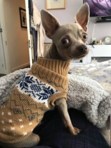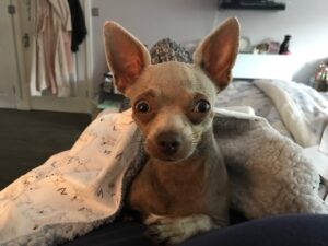
Boo is a one-year-old chihuahua who was referred to our dermatology clinic in August as he developed a non-inflammatory/non-pruritic alopecia in the spring. This mainly affected the ventrum and lower chest walls. The referring vet performed hair plucks and skin scrapings in July which were negative for Demodex mites. As the alopecia was slowly progressing, referral was arranged.
Examination
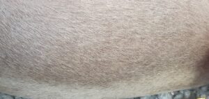
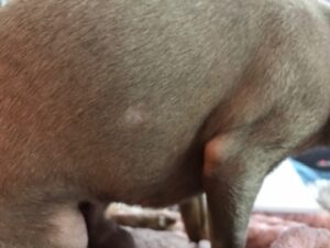
Boo was in good body condition and his weight was 2.6 kg. Physical examination was unremarkable. Dermatological examination revealed complete alopecia on the ventral chest and the lower part of both chest walls.
Almost complete alopecia was also seen on the ventrum and partial alopecia was seen affecting the groin as well as the upper thighs of both legs. Some comedones were seen around the testicles and upper thighs. The head, the dorsal neck, lower legs and tail were fully haired.
Differential diagnoses
Cause for partial (hypotrichosis) or complete alopecia would include:
· Congenital genetic abnormality (e.g. ectodermal defect)
· Folliculoadnexal inflammation (e.g. superficial pyoderma, demodicosis, dermatophytosis, sebaceous adenitis, alopecia areata, neoplasia)
· Trauma (e.g. pruritic skin disease)
· Hair cycling abnormalities (e.g. endocrinopathies, alopecia X, canine recurrent flank alopecia, defluxions)
· Malformed hair follicles (e.g. follicular dysplasia)
· Melanodysplasia (e.g. color-dilution alopecia, black hair follicular dysplasia)
· Disrupted blood supply (e.g. ischemic dermatopathy)
Diagnostic tests and diagnosis
We carried out a series of diagnostic tests to get to the root of Boo’s issues:
· Hair plucks and deep skin scrapings were taken to rule out demodicosis. No Demodex mites were found
· Trichogram showed most that most hairs were in the telogen phase. Melanosomes (melanin clumping) were present in some of the hair shafts
· Boo was sedated and four punch biopsies of 6 mm size were taken from the dorsal neck, right and left chest wall as well as upper thigh of right hind leg
· Histopathological analyses revealed follicular dysplasia with melanin clumping, consistent with colour dilution alopecia
Following these tests, the diagnosis was colour dilution alopecia.
Discussion and management
In this case, the clinicopathological correlation of the dermatological lesions and their distribution, the findings on the Trichogram, the breed and juvenile onset of clinical signs confirmed the diagnosis of colour dilution alopecia.
It is often forgotten that a Trichogram is a valuable diagnostic tool when dealing with cases of alopecia. This rapid and cost-effective test can aid to make a tentative diagnosis, or give an indication of the relevant differential diagnosis.
Trichogram allows to identify presence of ectoparasites such as Demodex mites or Louse nits or fungal elements (hyphae and arthrospores), as well as abnormalities of the hair bulb, the hair shaft and the hair tips. In Boo’s case, the presence of predominately telogen and melanin clumping supported the clinical suspicion of colour dilution alopecia and prompted to the next diagnostic step of taking skin biopsies.
Skin biopsies provide information about appearance of hair follicles as well as perifollicular tissue, presence of absence of sebaceous glands, presence of folliculoadnexal cellular infiltration as well the potential for hair re-growth through the existence anagen follicles. In this case skin biopsies were able to make a definitive diagnosis.
Colour dilution alopecia (CDA) – what is it?
Colour dilution alopecia (CDA) is the result of a genetically-based mutation of the dilution gene causing abnormal clumping of melanin within the hair shaft with subsequent fracturing and alopecia. However, other alleles and other factors may play an important role as well.
The disease usually does not manifest at birth. Age of onset is typically between three months to three years. It has been described in many canine breeds and is more prevalent in colour dilute individuals, especially those with a fawn (a dilution of a normally red or brown coat) or blue (a dilution of the normal black and tan colour).
Predisposed breeds include Doberman Pinschers, Yorkshire terriers, Great Danes, Whippets, Chihuahua, Dachshunds, Standard Poodles, Chow Chows and fawn Irish Setters. The patient’s fur usually looks normal at birth and then progresses gradually to develop a dry and dull coat with partial to complete alopecia on the dorsum of the trunk. Seborrhoea, comedones and secondary bacterial folliculitis often present in affected areas.
There is no cure for colour dilution alopecia but fortunately it is mainly a “cosmetic” disease and dogs can live a normal life. The condition can be managed by using medication to minimise scale and seborrhoea such as oral retinoids (vit A, isotretinoin, acitretin), supplements containing Omega 3 and six fatty acids, as well as epidermal barrier repair products such as Allerderm Spot-On, DOUXO S3 SEB Spot-on or Dermoscent Essential 6 pipettes. Medication to promote hair regrowth (e.g. melatonin) can also be used.
In this patient, a combination of oral melatonin, Omega 3 and Omega 6 supplement as well as Dermoscent essential 6 pipettes had led to clinical improvement within four weeks. The owner was advised to continue with the medication long term.