Tibial Plateau Levelling Osteotomy (TPLO) is a surgical procedure used to treat cranial (or anterior) cruciate ligament rupture in the knee joints of dogs.
Common causes for cruciate rupture
Chronic conditions:
- Degeneration (ageing) of the ligament
- Obesity/poor physical condition
- Skeletal shape/conformation of the dog
- Genetics
- Breed
Acute:
- Trauma such as twisting knee when jumping for a ball/toy
Pre-surgery a series of radiographs are needed. The surgeon will then use these radiographs to measure the correct angle and plate needed for a successfully surgery.
List of equipment
Radiography marker and position aids list:
- Left and right markers
- Ruler
- Radiographic reference ball OR radiographic measurer (used for all orthopaedic radiographs)
- Sandbags
- Blue wedges of different shapes and sizes
- Small foam blocks
- Swabs
- Ties (only when a patient is anaesthetised)
- Elastoplast (only when a patient is anaesthetised)
- Omnifilm tape
Pre-surgery radiographs
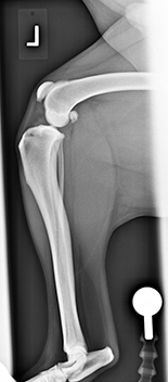
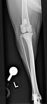
Lateral position
- Ensure that the stifle and hock are collimated into the area of interest. Please keep the leg in a natural position
- Place an appropriate-sized plate under the limb of interest
- Centre on stifle
- Collimate to include mid-femur and hock joint
- Sandbag the limb not being radiographed out of the way of the beam
- Use a blue flat triangular wedge under the hip/pelvis
- Left or Right marker
- Radiographic reference ball (place the ball at the level of the joint)
- You may or may not need a small foam block or some swabs to lay under the hock so it elevates the tibia/fibular parallel to the stifle
- Ensure the leg is in a natural position
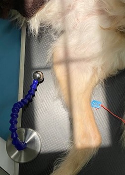
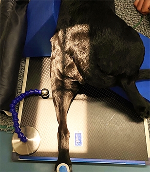
Caudocranial position
- Place patient on their front, hind limbs extended
- Place appropriate-sized plate under the limb of interest
- Centre on stifle
- Collimate to include all of mid femur and hock joint
- Place the non-interest limb onto wedges (this should help tilt the limb of interest into a more central position)
- Place a large blue triangular wedge/ big sandbag on the opposite side as this will help keep the patient in a central position
- Left or Right marker
- Radiographic reference ball (place the ball at the level of the joint)
- Use omnifilm to tape patient’s tail out of the way or tuck the tail under the non-interest limb if the tail is long enough
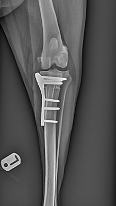
POST TPLO radiographs
Lateral position
- Ensure that the stifle and hock are collimated into the area of interest.
- Place appropriate sized plate under the limb of interest
- Centre on stifle
- Collimate to include mid femur and hock joint
- Sandbag the limb not being radiographed out of the way of the beam
- Blue flat triangular wedge under the hip/pelvis
Left or Right marker
- Radiographic reference ball (place the ball at the level of the joint)
- You may or may not need a small foam block or some swabs to lay under the hock so it elevates the tibia/fibular parallel to the stifle
These are guidelines and you may need more or less of the position aids as each patient will be different.